Last updated: April 23, 2013

Grabbing Hold Of Cells And Tissues With Zinc Fingers
Genome Advance of the Month
Grabbing hold of cells and tissues with zinc fingers
By Ian L. Marpuri
NHGRI Scientific Program Analyst
 |
This month's Genome Advance of the Month describes new uses for zinc fingers to improve researchers' ability to study the processes of single cells and interactions between larger groups of cells.
Prashant Mali, Ph.D., and John Aach, Ph.D., both from the lab of George Church, Ph.D., at Harvard Medical School studied the interactions of zinc fingers and DNA. They noticed that both zinc fingers and DNA are highly customizable, creating an endless number of combinations between them. This led them to explore the use of zinc fingers to create structures using cells and other large molecules.
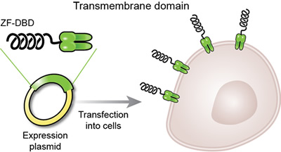
Their research team engineered zinc fingers that would be expressed on the surface of a living cell. These "surface zinc fingers" (sZFs) protrude out from the cell membrane so that DNA in the surrounding solution binds to them. The target DNA for each sZF can be fluorescently labeled so that any DNA binding to zinc fingers is visible under a special microscope. Since the zinc fingers show specific binding properties, they act as unique "barcodes" for the cells to which they are attached. Dr. Mali and his colleagues showed that mixtures of cells with different zinc finger labels can be isolated and visualized using fluorescently labeled DNA probes.
The team then devised a method to visualize large numbers of cell types using only three colors of fluorescent labels. After the first fluorescently labeled DNA probe binds to the zinc finger, two additional DNA probes bind to the first probe. The first of these two probes is a "quencher" that blocks the fluorescence from the initial probe. The second probe is fluorescently labeled with a different color. By watching the color transitions, six cells can be identified with only three colors. Repeating the sequence of quenching and adding a new fluorescent DNA probe allows for more and more cells to be visualized.
After successfully labeling cells, the team looked for other zinc finger applications.
The first potential function they found is using zinc fingers to report internal cell conditions. Researchers infected cells with DNA engineered to produce zinc fingers only in the presence of specific molecules. They looked for the presence of a molecule, such as a toxin, by looking for the surface zinc fingers rather than the toxin itself. They tested this with the antibiotic tetracycline and cumate, a small molecule, and easily detected the resulting zinc fingers. This application could help scientists study different molecular pathways by tracking when specific molecules are produced based on the appearance of zinc fingers.
The second use they considered is employing surface zinc fingers as molecular handles to capture specific cells. The researchers took a mixture of labeled and unlabeled cells and incubated them with magnetic beads covered in target DNA strands. After collecting the beads and washing them, the cells expressing zinc fingers were still attached to the beads. This would allow for easier isolation of particular cell types.
Third, the team showed that zinc fingers could help with the delivery of foreign DNA into cells. Scientists routinely use viruses to infect cells with foreign DNA for genetics research, but the success of that infection varies. These researchers showed that cells expressing zinc fingers were more easily infected by viruses coated in the zinc finger target DNA than by viruses without the target DNA. This could aid cell targeting, particularly when the cell of interest is mixed with other cell types.
But, the full potential of zinc fingers may not have been tapped yet.
"One intriguing potential use for this new method is to generate spatial arrangements of specific cell types to 'build' artificial tissues and organs," said Jeffery Schloss, Ph.D., director of NHGRI's division of genome science and program director for technology development coordination.
However, human tissue can be difficult to acquire and prepare since it requires a willing donor. Using zinc fingers, a scientist could create tissues for testing by combining individual cells. Scientists can produce the different cell types of a particular tissue or organ using induced pluripotent stem cells (iPSCs). These stem cells, produced from human tissue cells, can produce any other cell type. The different cell types would then get their own unique zinc finger barcode. Scientists could then create a molecular "paint by numbers" image by placing DNA probes where they want the specific cell types to anchor. This could be done on a flat surface or in a three dimensional framework.
"The resulting artificial tissue could be used for testing the effects of drug candidates," added Dr. Schloss.
This work represents a potentially big step in understanding how cells and tissues work and developing more accessible models for research. As this technique and other similar methods are developed, researchers may be able to generate tissues for use in research and in healthcare.