NHGRI logo

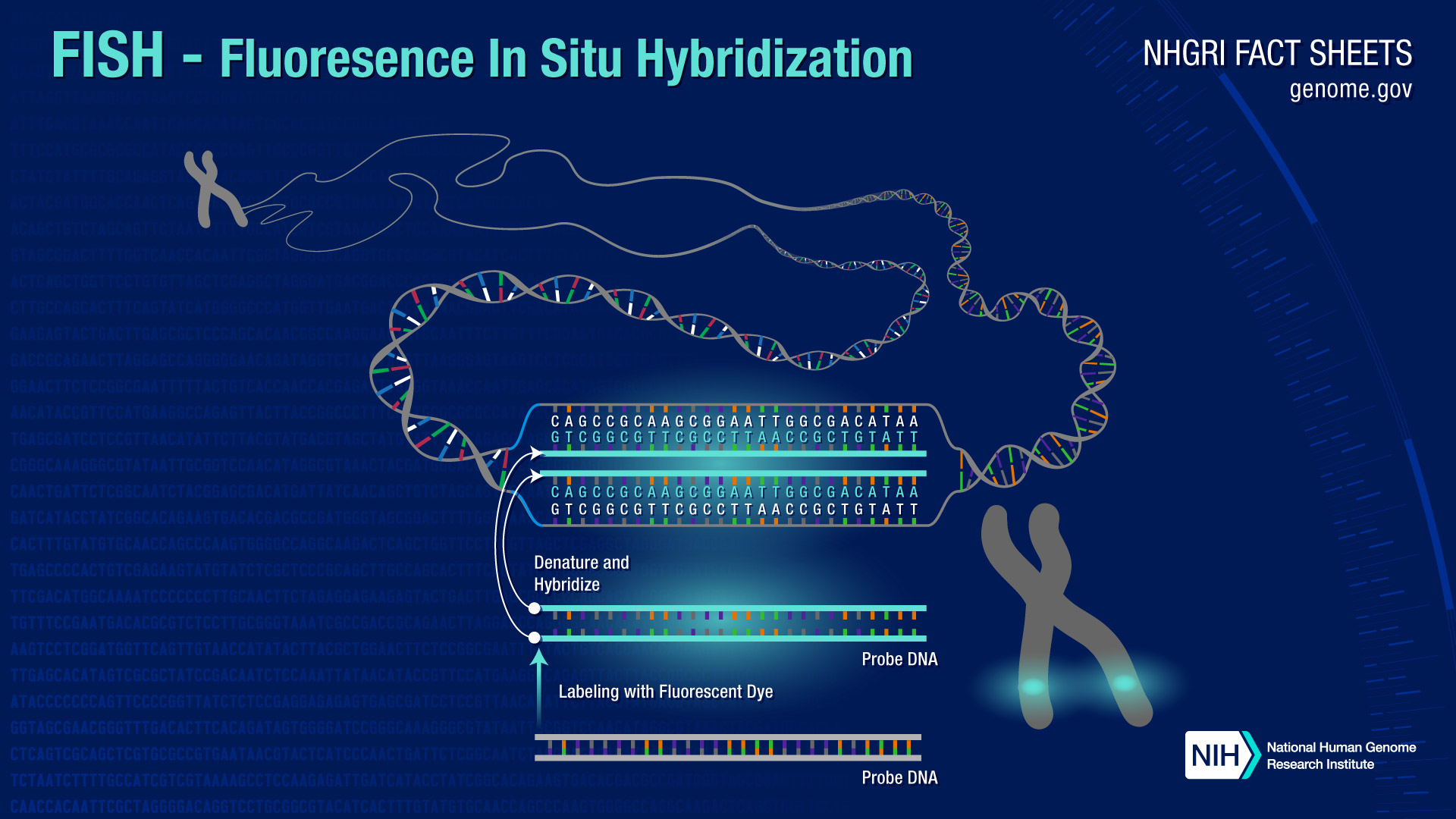
Fluorescence in situ hybridization (FISH) provides researchers with a way to visualize and map the genetic material in an individual's cells, including specific genes or portions of genes. This may be used for understanding a variety of chromosomal abnormalities and other genetic mutations.

How does FISH work?

FISH is useful, for example, to help a researcher or clinician identify where a particular gene falls within an individual's chromosomes. The first step is to prepare short sequences of single-stranded DNA that match a portion of the gene the researcher is looking for. These are called probes. The next step is to label these probes by attaching one of a number of colors of fluorescent dye.DNA is composed of two strands of complementary molecules that bind to each other like chemical magnets. Since the researchers' probes are single-stranded, they are able to bind to the complementary strand of DNA, wherever it may reside on a person's chromosomes. When a probe binds to a chromosome, its fluorescent tag provides a way for researchers to see its location.

  • How does FISH work?

    FISH is useful, for example, to help a researcher or clinician identify where a particular gene falls within an individual's chromosomes. The first step is to prepare short sequences of single-stranded DNA that match a portion of the gene the researcher is looking for. These are called probes. The next step is to label these probes by attaching one of a number of colors of fluorescent dye.DNA is composed of two strands of complementary molecules that bind to each other like chemical magnets. Since the researchers' probes are single-stranded, they are able to bind to the complementary strand of DNA, wherever it may reside on a person's chromosomes. When a probe binds to a chromosome, its fluorescent tag provides a way for researchers to see its location.

What is FISH used for?

Scientists use three different types of FISH probes, each of which has a different application:

Locus specific probes bind to a particular region of a chromosome. This type of probe is useful when scientists have isolated a small portion of a gene and want to determine on which chromosome the gene is located, or how many copies of a gene exist within a particular genome.

Alphoid or centromeric repeat probes are generated from repetitive sequences found in the middle of each chromosome. Researchers use these probes to determine whether an individual has the correct number of chromosomes. These probes can also be used in combination with "locus specific probes" to determine whether an individual is missing genetic material from a particular chromosome.

Whole chromosome probes are actually collections of smaller probes, each of which binds to a different sequence along the length of a given chromosome. Using multiple probes labeled with a mixture of different fluorescent dyes, scientists are able to label each chromosome in its own unique color. The resulting full-color map of the chromosome is known as a spectral karyotype. Whole chromosome probes are particularly useful for examining chromosomal abnormalities, for example, when a piece of one chromosome is attached to the end of another chromosome.

For many applications, FISH has largely been replaced by the use of microarrays. However, FISH remains useful for some tests. FISH may also be used to study comparisons among the chromosomal arrangements of genes across related species.


FISH

  • What is FISH used for?

    Scientists use three different types of FISH probes, each of which has a different application:

    Locus specific probes bind to a particular region of a chromosome. This type of probe is useful when scientists have isolated a small portion of a gene and want to determine on which chromosome the gene is located, or how many copies of a gene exist within a particular genome.

    Alphoid or centromeric repeat probes are generated from repetitive sequences found in the middle of each chromosome. Researchers use these probes to determine whether an individual has the correct number of chromosomes. These probes can also be used in combination with "locus specific probes" to determine whether an individual is missing genetic material from a particular chromosome.

    Whole chromosome probes are actually collections of smaller probes, each of which binds to a different sequence along the length of a given chromosome. Using multiple probes labeled with a mixture of different fluorescent dyes, scientists are able to label each chromosome in its own unique color. The resulting full-color map of the chromosome is known as a spectral karyotype. Whole chromosome probes are particularly useful for examining chromosomal abnormalities, for example, when a piece of one chromosome is attached to the end of another chromosome.

    For many applications, FISH has largely been replaced by the use of microarrays. However, FISH remains useful for some tests. FISH may also be used to study comparisons among the chromosomal arrangements of genes across related species.


    FISH

Last updated: August 16, 2020