
Chromosomes Fact Sheet
Chromosome Abnormalities Fact Sheet
Chromosome abnormalities can be numerical or structural. A numerical abnormality mean an individual is either missing one of the chromosomes from a pair or has more than two chromosomes instead of a pair. A structural abnormality means the chromosome's structure has been altered in one of several ways.
What are chromosomes?
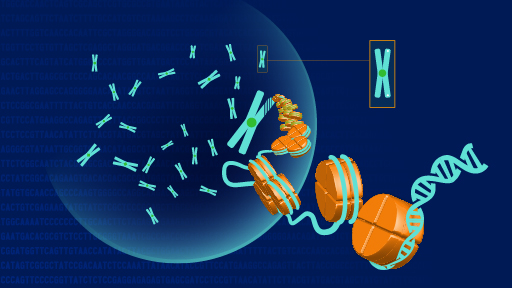
Chromosomes are the structures that hold genes. Genes are the individual instructions that tell our bodies how to develop and function; they govern physical and medical characteristics, such as hair color, blood type and susceptibility to disease.
Many chromosomes have two segments, called "arms," separated by a pinched region known as the centromere. The shorter arm is called the "p" arm. The longer arm is called the "q" arm.
-
What are chromosomes?
Chromosomes are the structures that hold genes. Genes are the individual instructions that tell our bodies how to develop and function; they govern physical and medical characteristics, such as hair color, blood type and susceptibility to disease.
Many chromosomes have two segments, called "arms," separated by a pinched region known as the centromere. The shorter arm is called the "p" arm. The longer arm is called the "q" arm.
Where are chromosomes found in the body?
The body is made up of individual units called cells. Your body has many different kinds of cells, such as skin cells, liver cells and blood cells. In the center of most cells is a structure called the nucleus. This is where chromosomes are located.
-
Where are chromosomes found in the body?
The body is made up of individual units called cells. Your body has many different kinds of cells, such as skin cells, liver cells and blood cells. In the center of most cells is a structure called the nucleus. This is where chromosomes are located.
How many chromosomes do humans have?
The typical number of chromosomes in a human cell is 46: 23 pairs, holding an estimated total of 20,000 to 25,000 genes. One set of 23 chromosomes is inherited from the biological mother (from the egg), and the other set is inherited from the biological father (from the sperm).
Of the 23 pairs of chromosomes, the first 22 pairs are called "autosomes." The final pair is called the "sex chromosomes." Sex chromosomes determine an individual's sex: females have two X chromosomes (XX), and males have an X and a Y chromosome (XY). The mother and father each contribute one set of 22 autosomes and one sex chromosome.
-
How many chromosomes do humans have?
The typical number of chromosomes in a human cell is 46: 23 pairs, holding an estimated total of 20,000 to 25,000 genes. One set of 23 chromosomes is inherited from the biological mother (from the egg), and the other set is inherited from the biological father (from the sperm).
Of the 23 pairs of chromosomes, the first 22 pairs are called "autosomes." The final pair is called the "sex chromosomes." Sex chromosomes determine an individual's sex: females have two X chromosomes (XX), and males have an X and a Y chromosome (XY). The mother and father each contribute one set of 22 autosomes and one sex chromosome.
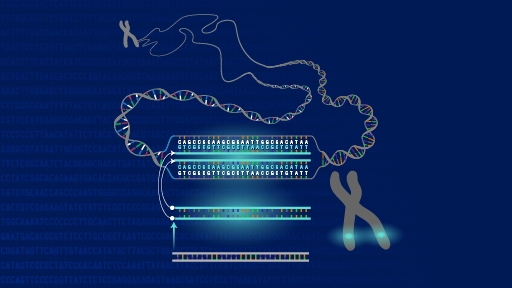
How do scientists study chromosomes?
For a century, scientists studied chromosomes by looking at them under a microscope. In order for chromosomes to be seen this way, they need to be stained. Once stained, the chromosomes look like strings with light and dark "bands," and their picture can be taken. A picture, or chromosome map, of all 46 chromosomes is called a karyotype. The karyotype can help identify abnormalities in the structure or the number of chromosomes.
To help identify chromosomes, the pairs have been numbered from 1 to 22, with the 23rd pair labeled "X" and "Y." In addition, the bands that appear after staining are numbered; the higher the number, the farther that area is from the centromere.
In the past decade, newer techniques have been developed that allow scientists and doctors to screen for chromosomal abnormalities without using a microscope. These newer methods compare the patient's DNA to a normal DNA sample. The comparison can be used to find chromosomal abnormalities where the two samples differ.
One such method is called noninvasive prenatal testing. This is a test to screen a pregnancy to determine whether a baby has an increased chance of having specific chromosome disorders. The test examines the baby's DNA in the mother's blood.
-
How do scientists study chromosomes?
For a century, scientists studied chromosomes by looking at them under a microscope. In order for chromosomes to be seen this way, they need to be stained. Once stained, the chromosomes look like strings with light and dark "bands," and their picture can be taken. A picture, or chromosome map, of all 46 chromosomes is called a karyotype. The karyotype can help identify abnormalities in the structure or the number of chromosomes.
To help identify chromosomes, the pairs have been numbered from 1 to 22, with the 23rd pair labeled "X" and "Y." In addition, the bands that appear after staining are numbered; the higher the number, the farther that area is from the centromere.
In the past decade, newer techniques have been developed that allow scientists and doctors to screen for chromosomal abnormalities without using a microscope. These newer methods compare the patient's DNA to a normal DNA sample. The comparison can be used to find chromosomal abnormalities where the two samples differ.
One such method is called noninvasive prenatal testing. This is a test to screen a pregnancy to determine whether a baby has an increased chance of having specific chromosome disorders. The test examines the baby's DNA in the mother's blood.
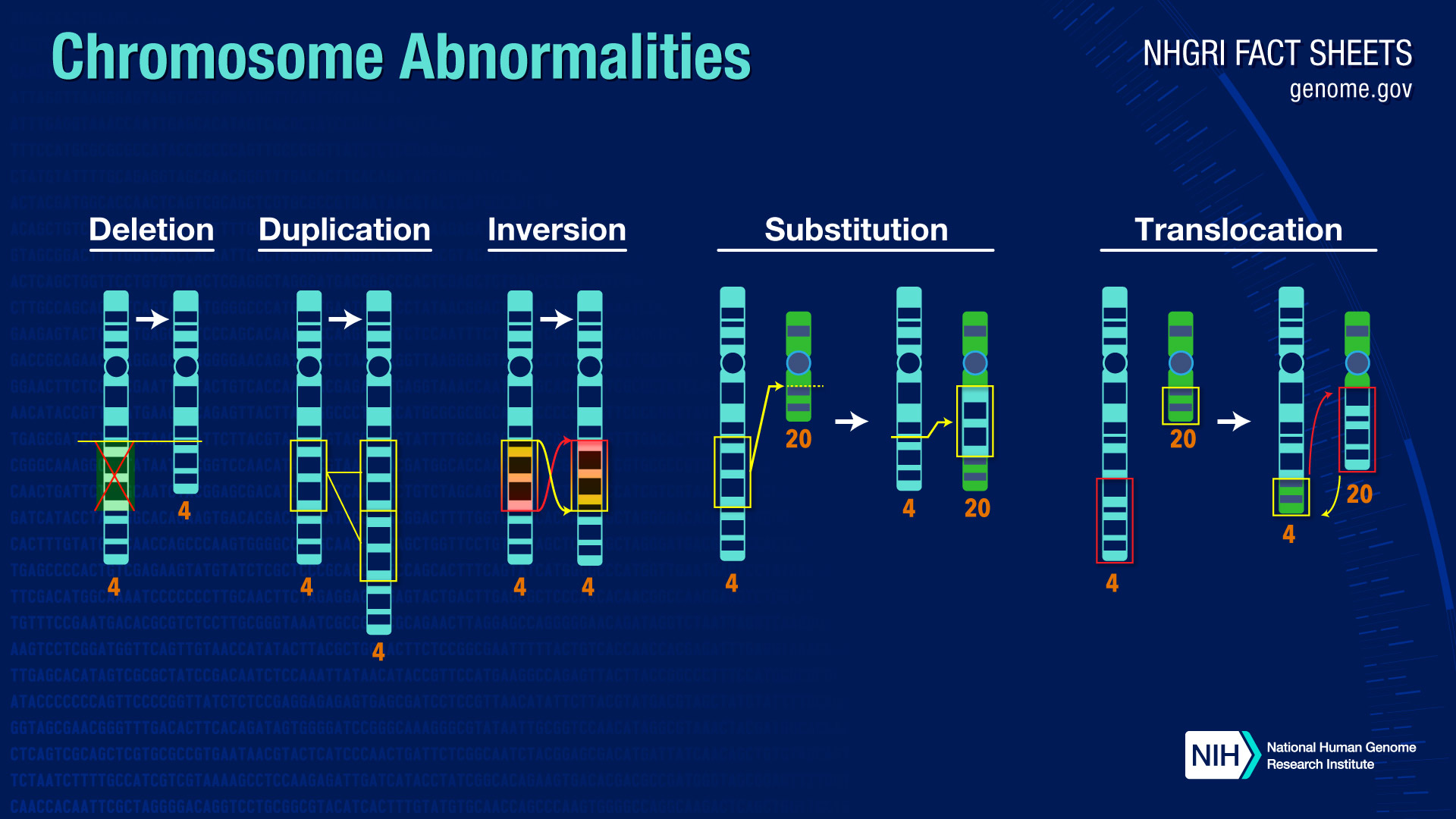
What are chromosome abnormalities?
There are many types of chromosome abnormalities. However, they can be organized into two basic groups: numerical abnormalities and structural abnormalities.
-
Numerical Abnormalities: When an individual is missing one of the chromosomes from a pair, the condition is called monosomy. When an individual has more than two chromosomes instead of a pair, the condition is called trisomy.
An example of a condition caused by numerical abnormalities is Down syndrome, which is marked by mental disability, learning difficulties, a characteristic facial appearance and poor muscle tone (hypotonia) in infancy. An individual with Down syndrome has three copies of chromosome 21 rather than two; for that reason, the condition is also known as Trisomy 21. An example of monosomy, in which an individual lacks a chromosome, is Turner syndrome. In Turner syndrome, a female is born with only one sex chromosome, an X, and is usually shorter than average and unable to have children, among other difficulties.
-
Structural Abnormalities: A chromosome's structure can be altered in several ways.
-
Deletions: A portion of the chromosome is missing or deleted.
-
Duplications: A portion of the chromosome is duplicated, resulting in extra genetic material.
-
Translocations: A portion of one chromosome is transferred to another chromosome. There are two main types of translocation. In a reciprocal translocation, segments from two different chromosomes have been exchanged. In a Robertsonian translocation, an entire chromosome has attached to another at the centromere.
-
Inversions: A portion of the chromosome has broken off, turned upside down, and reattached. As a result, the genetic material is inverted.
-
Rings: A portion of a chromosome has broken off and formed a circle or ring. This can happen with or without loss of genetic material.
-
Most chromosome abnormalities occur as an accident in the egg or sperm. In these cases, the abnormality is present in every cell of the body. Some abnormalities, however, happen after conception; then some cells have the abnormality and some do not.
Chromosome abnormalities can be inherited from a parent (such as a translocation) or be "de novo" (new to the individual). This is why, when a child is found to have an abnormality, chromosome studies are often performed on the parents.
-
How do chromosome abnormalities happen?
Chromosome abnormalities usually occur when there is an error in cell division. There are two kinds of cell division, mitosis and meiosis.
-
Mitosis results in two cells that are duplicates of the original cell. One cell with 46 chromosomes divides and becomes two cells with 46 chromosomes each. This kind of cell division occurs throughout the body, except in the reproductive organs. This is the way most of the cells that make up our body are made and replaced.
-
Meiosis results in cells with half the number of chromosomes, 23, instead of the normal 46. This is the type of cell division that occurs in the reproductive organs, resulting in the eggs and sperm.
In both processes, the correct number of chromosomes is supposed to end up in the resulting cells. However, errors in cell division can result in cells with too few or too many copies of a chromosome. Errors can also occur when the chromosomes are being duplicated.
Other factors that can increase the risk of chromosome abnormalities are:
-
Maternal Age: Women are born with all the eggs they will ever have. Some researchers believe that errors can crop up in the eggs' genetic material as they age. Older women are at higher risk of giving birth to babies with chromosome abnormalities than younger women. Because men produce new sperm throughout their lives, paternal age does not increase risk of chromosome abnormalities.
-
Environment: Although there is no conclusive evidence that specific environmental factors cause chromosome abnormalities, it is still possible that the environment may play a role in the occurrence of genetic errors.
-
Related Contents

Last updated: August 15, 2020